
Introduction to the Anatomy of the Wrists and Hands
Grip strength depends on the health and function of the structures of the hand, wrist, and forearm. If you understand the anatomy of these structures, you’ll be better able to take action to protect them.
Before we start, take a few minutes to perform a task you might do several times a week. Go to your clothes closet, open the door, take a shirt or blouse (with buttons) off of a hangar, put it on, and button it up. If your arms and hands are healthy and pain-free, this should have been an easy task.
Now, put the shirt back, close the closet door, and start the task all over again. This time, though, pay close attention to the complex movements of your wrist and hand that occur as you repeat these tasks. Were you amazed at all the movements that occurred and the variety of positions required of your forearms, wrists, and hands?
The Anatomical Position
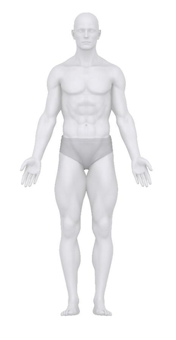
When we discuss the structures of the hand, it will help if you know how the arms and hands are positioned in the anatomical position. This is the position of the body that health care professionals use when describing movements such as flexion (bending) and extension (straightening).
When you look at the graphic below, you can see that the forearms are rotated so that the model’s palms are facing outward. When we talk about movements of the hand and wrist, go back to this graphic for your frame of reference.
The Bones of the Forearms, Wrists, and Hands
Take a look at the image below. It shows all the bones of the upper extremity. The bones serve as a framework for the muscles to pull on to allow for movement of the wrist and hand. Each arm (above the elbow) contains just one bone—the humerus. Each forearm (below the elbow) contains just two bones—the radius and the ulna.

Source: Shutterstock. Used with permission.
Each wrist and hand, however, contains 27 bones—an architectural marvel. The bones of the wrist include the eight bones called the carpals. Your grip strength depends on the carpals’ ability to create stability in the wrist while allowing subtle gliding movements between them. The bones of your fingers are called the phalanges. Each finger has three phalanges, except for the thumb which has only two.
The Joints of the Wrists and Hands
Hand function also depends on how freely the joints between bones move. Joints form the junctions between bones and allow for movement. Besides the movement between the carpals, the bottom row of carpals forms joints with the bones of the forearm—the radius and ulna. The top row of carpals forms joints with the bones that make up the flat part of your hand—the metacarpals. The metacarpals form joints between the first row of bones in the fingers—the phalanges, and the joints between your phalanges allow you to bend and straighten your fingers.
A smooth substance called cartilage covers the ends of the bones in the wrist and hand. This cartilage can get damaged by overuse, injury, or the effects of aging. If damage to the cartilage occurs, osteoarthritis may result, causing pain and stiffness. You can read more about osteoarthritis in the blog on aging and the hand.

Source: Shutterstock. Used with permission.
The Ligaments of the Hands
Ligaments link bones together. The image below shows you just some of the ligaments that connect the eight carpal bones in the wrist. You can also see some of the ligaments of the hand as well. Injuries to these ligaments will affect the function of your hands, including your grip strength.

Source: Shutterstock. Used with permission
Tough connective tissue makes up the ligaments of your hands. While this tissue is tough, injuries to the ligaments of the hands and wrists occur fairly often. Unfortunately, ligaments don’t contain a good blood supply, so when they are injured, healing can be difficult.
Doctors and therapists call an injury to a ligament a sprain. Because ligaments allow your hands to move, while providing stability, an injury to one or more ligaments can seriously decrease the ability of your hand and wrist to move properly. When this happens, everyday tasks, like putting on a shirt or blouse, can become difficult or impossible.
If any of the joints in your hands and wrists become painful, swollen, or difficult to move, you should consult a hand specialist right away to avoid further damage and permanent disability.
Muscles that Control the Wrists and Hands
You already know that each hand and wrist contains 27 bones. The number of muscles that control the wrists and hands is also impressive—each hand and wrist is controlled by 34 muscles. We won’t discuss all the muscles—just enough to give you an idea of how they work together to allow for all the complicated movements the hands and wrists perform.
If you’re sitting down at a desk, rotate your right forearm so that your palm is faced upward. Therapists call this movement supination and, while it appears that the movement occurs at the wrist, the movement is really occurring at the elbow. The images below show you a right forearm, wrist, and hand that’s in the anatomical position with the forearm supinated.
The superficial muscles lie just underneath the skin, the deep muscles lie closest to the bone, and the middle muscles are in between them.

Source: Shutterstock. Used with permission.
Let’s take a look at some of these muscles and their actions.
- Palmaris Longus Muscle: The palmaris longus muscle flexes the wrist. You can see from the image above that its tendon inserts into tissue on top of the wrist bones (the carpals). Tendons usually attach muscles to bones. The palmaris longus, however, attaches to a band of connective tissue that circles the wrist called the retinaculum and tissue found at the base of the palm.

Flexion of wrist with fingers extended
Photo credit: Morris Attaway. Used with permission.
- The flexor carpi radialis muscle and the flexor digitorum superficialis muscle: These two superficial muscles flex both the wrist and the fingers.
- The flexor digitorum profundus muscle and the flexor pollicis longus muscle: These two deep muscles also flex both the wrist and the fingers.
The photograph below shows you the action of these muscles.

Flexion of wrist, fingers and thumb
Photo credit: Morris Attaway. Used with permission.
Now rotate your right forearm so that your palm is resting on the table in a pronated position. The images below show you the muscles of your hand, wrist and forearm when your right forearm is pronated.

Source: Shutterstock. Used with permission.
Let’s take a look at some of these muscles and their actions:
- Extensor carpi radialis longus, extensor carpi radialis brevis, and extensor carpi ulnaris muscles: Because these muscles attach to the base of the metacarpal bones, they extend the wrist without extending the fingers.
- Extensor digitorum and the extensor pollicis muscles: These muscles extend the wrist while extending the fingers.
The photograph below shows all these muscles working together to extend both the wrist and the fingers.

Extension of wrist and fingers
Photo credit: Morris Attaway. Used with permission.
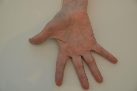
Let’s look now at muscles that stay entirely within the hands. The image below shows you these muscles as well as the tendons of some of the muscles that flex the wrist and fingers. You are looking at a right hand, with the palm turned up (supinated).

Source: Shutterstock. Used with permission
The muscles that stay inside the hands (the intrinsic hand muscles) form a complicated system of muscles. All you need to know is that some of these muscles spread the fingers apart (finger abduction), some of them bring the fingers closer together (finger adduction), and some allow you to flex the joints between the metacarpals and phalanges without flexing the fingers.
Here are three photographs that will illustrate these movements:
Finger and Thumb Abduction
Photo credit: Morris Attaway. Used with permission.

Finger and Thumb Adduction
Photo credit: Morris Attaway. Used with permission.

The intrinsic muscles of the hand working together
Photo credit: Holly Trimble. Used with permission.
The Nervous System’s Role
A discussion of wrist and hand function wouldn’t be complete without mention of the importance of the nervous system. Your brain receives information about the exact position of all the structures of your hands. It also sends messages to the muscles of your hands to tell them exactly when to move and with how much force.
Here’s an image that shows you how much brain power is devoted to receiving sensations from your hands. A similar amount of brain power is devoted to directing the movement of the structures of the hands.

Source: Shutterstock. Used with permission.
We just touched on the anatomy and movements of the hands and wrists in this article. This great resource will give you more information about these structures if you want to deepen your understanding of their anatomy and function.
Disclaimer: This article was written for educational purposes only. Any concerns about the health of your wrists and hands should be brought to the attention of your doctor or a hand specialist.
About the Author
Holly Trimble, PT, DPT earned three degrees in physical therapy, including a doctorate, and a master’s degree in biology. She worked as a physical therapist for fifteen years and taught college-level anatomy and physiology for sixteen years. Dr. Trimble currently writes and teaches health and science courses for Ed2go, Inc.


Share:
Grip Strength and Cardiovascular Health
Common Causes of Wrist and Hand Weakness